Diabetic Retinopathy
If you have diabetes mellitus, your body does not use and store sugar properly. Diabetes can cause changes in the body's blood vessels, the veins and arteries that carry blood throughout your body. Together with high blood pressure, it can affect vision by causing cataracts, glaucoma and, most importantly, damage to blood vessels inside the eye.
What is Diabetic and Hypertensive Retinopathy?
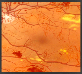
Retinopathy is a complication of diabetes and hypertension that is caused by changes in the blood vessels of the eye. The retina is a nerve layer at the back of the eye that senses light and helps to send images to your brain. When blood vessels in the retina are damaged, they may leak fluid or blood, and grow fragile, brush-like branches and scar tissue. This can blur or distort the images that the retina sends to the brain.
Diabetic retinopathy is the leading cause of new blindness among adults in the United States. People with untreated diabetes are said to be 25 times more at risk for blindness than the general population. The longer a person has diabetes and hypertension, the more the risk of developing retinopathy increases. About 80% of the people who have had diabetes for at least 15 years have some blood vessel damage to their retina. People with Type I, or juvenile, diabetes are more likely to develop diabetic retinopathy at a younger age.
If you have diabetes or hypertension, it's important to know that today, with improved methods of diagnosis and treatment, only a small percentage of people who develop retinopathy have serious vision problems.
Types of Diabetic Retinopathy
Background or non-proliferative retinopathy is an early stage of diabetic retinopathy. In this stage, tiny blood vessels within the retina become damaged and leak blood or fluid. Leaking fluid causes the retina to swell or to form deposits called exudates. While this stage usually doesn't affect your vision, it can lead to more sight threatening stages. For this reason, background retinopathy is considered a warning sign. Sometimes the leaking fluid collects in the macula, the part of the retina that lets us see fine details, like letters or numbers. This problem is called macular edema. Reading and close work may become more difficult because of this condition.
Proliferative retinopathy describes the changes that occur when new, abnormal blood vessels begin growing on the surface of the retina. The abnormal growth is called neovascularization. These new blood vessels have weaker walls and may break and bleed. The vitreous is the clear, jelly-like substance that fills the center of the eye. Leaking blood can cloud the vitreous and partially block the light passing through the pupil towards the retina, causing blurred and distorted images. These abnormal blood vessels may grow scar tissue that can pull the retina away from the back of the eye. This is called a retinal detachment. If left untreated, a retinal detachment can cause severe vision loss. Abnormal blood vessels may also grow around the pupil (on the iris) causing glaucoma by increasing pressure within the eye. Proliferative diabetic retinopathy is the most serious form of diabetic retinal disease. It affects up to 20% of diabetics and can cause severe loss of sight, including blindness.
Proliferative retinopathy describes the changes that occur when new, abnormal blood vessels begin growing on the surface of the retina. The abnormal growth is called neovascularization. These new blood vessels have weaker walls and may break and bleed. The vitreous is the clear, jelly-like substance that fills the center of the eye. Leaking blood can cloud the vitreous and partially block the light passing through the pupil towards the retina, causing blurred and distorted images. These abnormal blood vessels may grow scar tissue that can pull the retina away from the back of the eye. This is called a retinal detachment. If left untreated, a retinal detachment can cause severe vision loss. Abnormal blood vessels may also grow around the pupil (on the iris) causing glaucoma by increasing pressure within the eye. Proliferative diabetic retinopathy is the most serious form of diabetic retinal disease. It affects up to 20% of diabetics and can cause severe loss of sight, including blindness.
What Are the Symptoms of Diabetic Retinopathy?
There are usually no symptoms of background retinopathy, although gradual blurring of vision may occur if macular edema is present. You may never notice changes in your vision. An ophthalmic examination is the only way to find changes inside your eye. When bleeding occurs, your sight may become hazy, spotty or even disappear altogether. While there is no pain, proliferative retinopathy is a severe form of the disease and requires immediate medical attention. Pregnancy and high blood pressure may aggravate diabetic retinopathy
How Is Diabetic Retinopathy Diagnosed?
The best protection against diabetic retinopathy is to have regular medical eye examinations by your ophthalmologist. Serious retinopathy can be present without any symptoms. The disease can improve with treatment. To find diabetic retinopathy, the ophthalmologist looks at the inside of the eye using an instrument called an ophthalmoscope. The pupils may need to be dilated (enlarged) with eye drops. If your ophthalmologist finds diabetic retinopathy, he or she may order color photographs of the retina or a special test called fluorescein angiography to find out if you need treatment. Fluorescein angiography is a test where dye is injected in your arm and special photos of your eye are taken.
How Is Diabetic Retinopathy Treated?
Your Ophthalmologist Will Consider:
In advanced proliferative diabetic retinopathy, the ophthalmologist may recommend intraocular injections or a vitrectomy. This microsurgical procedure is performed in the operating room. Vitrectomy removes the blood-filled vitreous and replaces it with a clear solution. About 70% of vitrectomy patients notice an improvement in sight after surgery. Sometimes the ophthalmologist may wait from several months up to a year to see if the blood clears on its own, before going ahead with a vitrectomy.
- Your age
- Your medical history
- Your lifestyle
- How much your retina is damaged
In advanced proliferative diabetic retinopathy, the ophthalmologist may recommend intraocular injections or a vitrectomy. This microsurgical procedure is performed in the operating room. Vitrectomy removes the blood-filled vitreous and replaces it with a clear solution. About 70% of vitrectomy patients notice an improvement in sight after surgery. Sometimes the ophthalmologist may wait from several months up to a year to see if the blood clears on its own, before going ahead with a vitrectomy.
Diabetic retinopathy is the leading cause of new blindness among adults in the United States. People with untreated diabetes are said to be 25 times more at risk for blindness than the general population.
Vision Loss Is Largely Preventable
Diabetic retinopathy may be present without any symptoms. Early detection of diabetic retinopathy is the best protection against loss of vision. People with diabetes should schedule examinations by an ophthalmologist at least once a year. More frequent medical eye examinations may be necessary once diabetic retinopathy has been diagnosed. With careful monitoring, the ophthalmologist can begin treatment before sight is affected. When indicated, laser, medical and operative surgery are highly effective treatments for diabetic retinopathy.